- Mon - Sat
08:00am - 08:00pm


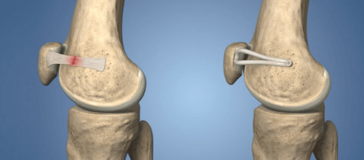
MEDIAL PATELLO FEMORAL LIGAMENT (MPFL), attaches to the inner side of patella (knee cap) to the long bone of our thigh (femur). It provides stability to patella from inner side, so during knee flexion and extension movement of knee, patella glides in the trochlear notch of femur smoothly.
MPFL along with other muscles and ligamants functions to keep track of patella in the trochlear groove and prevents it from dislocating / subluxating.
MPFL can be injured due to direct trauma or can tear / stretch following lateral dislocation of patella (knee cap) from trochlear groove. MPFL can also be lax as a result of generalised congenital ligamentous laxity.
Commonest is traumatic dislocation of patella. MPFL insufficiency is often associated with an anatomic abnormality in knee leading to patellar dislocation.
Traumatic first time dislocation of patella is usually treated conservatively with RICE and physiotherapy, but recurrent dislocation needs surgical treatment. Left untreated repeated patellar dislocation can cause cartilage damage and early arthritic changes in the knee.
The orthopedist at AOSM after careful exam will investigate often by Xray and MRI and look for the MPFL tear.
If there is isolated MPFL tear than reconstruction of MPFL is treatment of choice.
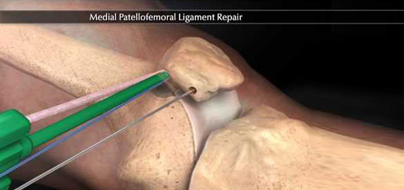
MPFL reconstruction is a procedure wherein surgeon harvests graft (tendon) from one of the hamstring muscle, and reconstructs the ligament by attaching graft from inner side of patella to femur bone. Surgeon makes two small incision in the knee and then he creates plane between inner side of patella to femur, graft is passed through a tunnel in patella and fixed to femur bone with a screw in desired position. Post operatively knee is immobilised in a long knee brace for 3-6 weeks. Early rehab is started and usually patient is back to most activity by 3 months.
